Overview
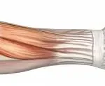
The saphenous nerve is the longest purely sensory branch of the femoral nerve, arising primarily from the L3–L4 nerve roots. It travels down the inner thigh, crosses the knee region, and continues along the medial side of the lower leg to the area just above the ankle. Its primary role is to transmit sensory information, touch, pain, and temperature from the skin on the inner thigh, medial knee, and inner aspect of the leg and ankle to the spinal cord. Because it does not supply any muscles, the saphenous nerve carries no motor fibres.
Saphenous nerve entrapment describes a situation in which this nerve becomes compressed, stretched, or otherwise irritated as it courses through soft tissue structures. The entrapment can occur at several anatomical points, but the most common location is the adductor (subsartorial) canal, a passage beneath the sartorius muscle bordered by fascia and other structures. Here, tight or hypertrophied muscles, thickened fibrous bands, surgical scarring, or even minor trauma can reduce the space available for the nerve, leading to its compression. Although the saphenous nerve does not control muscles, disruption of its sensory signalling can still cause significant discomfort, numbness, or altered sensation along its distribution.
Within the context of clinics such as DMPhysios in Noida, renowned for its focus on spine and sports conditions and its patient-centred rehabilitation approach, saphenous nerve entrapment represents a frequently overlooked but clinically important cause of medial knee pain, inner leg hypersensitivity, or unusual sensations below the knee. Identifying the condition early and implementing a well-designed physiotherapy programme can greatly improve outcomes, reduce chronic pain, and help patients return to normal activity more quickly.
Symptoms
The clinical presentation of saphenous nerve entrapment can vary depending on the site of entrapment and severity. Common symptoms include:
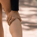
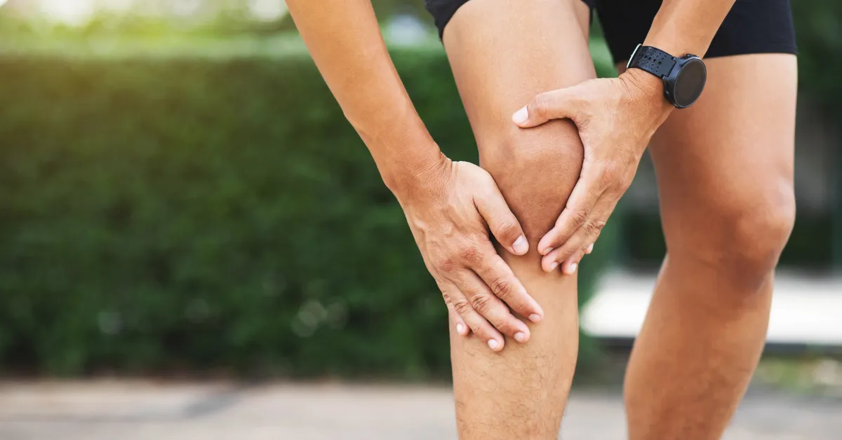
- Pain: Often described as burning, aching, or sharp in the inner knee, medial thigh, inner lower leg, sometimes down to the ankle. Aggravated by activities such as walking, stair climbing, knee extension, or sustained positions.
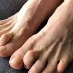
- Paresthesia: Tingling, numbness, or “pins and needles” sensations in the sensory distribution of the saphenous nerve (inner thigh, inner knee, medial lower leg, inner ankle).
- Sensitivity to touch: Light touch or pressure over certain points (e.g. adductor canal, medial femoral condyle) may provoke symptoms.
- Increased discomfort with certain movements: Knee flexion, hip extension, abduction, or compression at the adductor canal. Sometimes pain worsens at night.
- No motor deficits: Because the saphenous nerve is sensory only, motor strength is preserved. If there is weakness, consider other nerves or spinal root involvement.
Types & Sites of Entrapment
There are different anatomical variants and branch-specific entrapments. Some “types” based on location:
- Adductor canal (subsartorial canal) entrapment
The saphenous nerve passes through the adductor canal; entrapment here is common and may involve compression by fibrous bands or vascular structures. - Infrapatellar branch entrapment
The infrapatellar branch (branch of the saphenous nerve) crosses the knee region, sometimes through or under the sartorius, or through fascial layers. It can become entrapped separately. - Branch entrapment in sartorius tendon / beneath muscular / fascial layers
For example, the infrapatellar branch may pierce the sartorius, or run under/through fascia in ways that make it vulnerable. - Multiple compression points
Sometimes more than one site of entrapment exists along the course of the saphenous nerve.
These types are important because location determines what symptoms are dominant, what provocative tests will reproduce the symptoms, and what interventions are most effective.
Causes
What causes saphenous nerve entrapment ? Some of the key etiologies:
- Anatomical/fascial constraints: Tight or hypertrophic sartorius muscle, fibrous bands, the vastoadductor membrane, fascia through which branches pass.
- Surgical trauma / iatrogenic injury: Knee arthroscopy, ACL reconstruction (hamstring graft harvest), total knee arthroplasty, incisions along the medial knee, varicose vein surgery, or surgeries involving the adductor canal.
- Scar tissue / neuroma formation: Post-surgical or post-traumatic scar tissue can trap the nerve or its branches. Neuroma of the infrapatellar branch is one such cause.
- Trauma / direct blow: Violence, contusion, or repetitive stress on the medial knee or thigh.
- Compression from surrounding structures: Vessels, tight clothes or braces, swelling, varicosities, or masses (rarely tumours).
- Overuse / sports stress: Running, repetitive movements that stretch or compress the adductor canal, or pushing the knee into certain ranges repeatedly.
Risk Factors
These are the conditions or traits that increase likelihood of saphenous nerve entrapment :
- Prior knee surgery (especially involving medial incisions) or trauma.
- Sports with high mileage or frequent repetitive stress on knee / inner thigh (runners, ultramarathoners)
- Anatomical variation (nerve piercing sartorius, unusual fascial attachments) that predispose to compression.
- Excessive tightness of muscles surrounding the adductor canal (sartorius, adductors) or poor flexibility.
- Scarring from surgeries, injury or inflammation.
- Aging and degenerative changes which might affect fascial elasticity or soft tissue compliance.
- Obesity or external compression (tight garments, braces, etc.) which increase pressure around the nerve course.
Diagnosis
While you didn’t ask explicitly for diagnosis, it’s relevant for understanding treatment. Common diagnostic steps:
- History & physical exam focusing on sensory symptoms without motor loss; palpation of adductor canal; provocative movements (hip extension, abduction and knee flexion may stretch nerve)
- Diagnostic nerve block (local anesthetic injection) in the adductor canal to see if symptoms improve.
- Imaging: MRI or ultrasound to look for masses, nerve relationship to muscles/fascia.
- Electrophysiological studies (nerve conduction / EMG) are less useful since saphenous is purely sensory, but sometimes helpful to rule out other causes. Also may be negative even when entrapment is present.
Treatment
Treatment of saphenous nerve entrapment generally proceeds from conservative to more invasive, depending on duration, severity, and response. Key options include:
- Rest & activity modification – avoiding aggravating activities, positions, or pressure.
- Pain control – NSAIDs, modalities like heat/ice, possibly local corticosteroid injection.
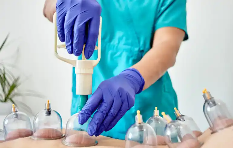
- Manual therapies – soft tissue mobilization, fascia release.
- Neural mobilisation / nerve gliding or flossing techniques.
- Physical therapy / rehabilitation (see more under physiotherapy treatment).
- If conservative fails, surgical decompression or release of the entrapped branch. For example, partial division of sartorius in infrapatellar branch entrapment.
Physiotherapy Treatment
At DMPhysios, which focuses on patient-centred rehabilitation for spine and sports conditions, physiotherapy for saphenous nerve entrapment can be highly effective when tailored to the individual. Below is a detailed physiotherapy protocol / plan.
Assessment
- Detailed subjective history: onset, aggravating/relieving factors, duration, prior surgeries.
- Mapping of sensory disturbance: inner thigh, knee, lower leg.
- Palpation of adductor canal, medial knee region, infra-patellar branch path.
- Provocative tests: stretching (hip extension + abduction + knee flexion), compression over suspected entrapment site.
- Assess muscle tightness (sartorius, adductors, quadriceps medial side), joint range of motion (hip, knee).
- Evaluate posture, gait, biomechanics (e.g. knee alignment, foot pronation), and overall lower limb strength.
Treatment Phases
- Acute Phase (Pain control, decreasing nerve irritability)
- Relative rest: avoid activities that stretch or compress the saphenous nerve.
- Pain modulation modalities: heat, ice, ultrasound or electrical stimulation as tolerated.
- Gentle soft tissue mobilization around the area of entrapment (adductor canal, sartorius) to reduce fascial tightness.
- Gentle stretching of adjacent muscles: adductors, sartorius, hip flexors.
- Mobilization / Neural mobilisation Phase
- Nerve gliding / flossing exercises for the saphenous nerve entrapment. These are controlled exercises that allow the nerve to gently slide through its anatomical pathways without exacerbating symptoms. Over time, gliding may help reduce adhesions and improve mobility.
- Soft tissue release / myofascial techniques: focusing on fascia around sartorius, medial knee, adductor canal.
- Strengthening / Biomechanical Correction
- Strengthen hip abductors, external rotators, core stabilization to correct biomechanical stresses that contribute to entrapment.
- Quadriceps strengthening, especially VMO, ensures symmetric loading.
- Adductor strengthening, but carefully, so as not to overcompress adductor canal early in rehab.
- Flexibility & Mobility
- Stretching of hip adductors, sartorius, hamstrings, quadriceps medial side.
- Joint mobilizations if there is stiffness in hip or knee joints contributing to altered movement patterns.
- Functional & Sports-Specific Conditioning
- Gradual return to weight-bearing / dynamic activities (walking, running, stair climbing) with modulation of load.
- Movement retraining: gait, landing mechanics, knee alignment.
- Education in activity pacing, use of supportive devices if needed temporarily (e.g., taping, protective gear) to avoid external compression.
- Monitoring & Adjustment
- Reassessment regularly: pain levels, sensory symptoms, functional measures.
- Adjust interventions based on response. If symptoms persist beyond a reasonable conservative period (4-12 weeks depending on severity), consider referral for further investigation or surgical consultation.
Specific Physiotherapy Techniques
- Soft Tissue Release (STR) around the sartorius muscle and adductor region to reduce compression.
- Trigger Point Release in adjacent musculature that may be contributing to tightness.
- Neurodynamic Techniques / Nerve Mobilizations, specific to saphenous nerves (gliding vs gentle tension depending on patient tolerance).
- Scar Tissue Mobilization if present (post-surgical).
- Modalities: low-level laser, ultrasound, TENS if pain is high.
At DMPhysios, patient-centred rehab will also include educating the patient about their condition of saphenous nerve entrapment, self-management strategies (home exercise, posture / movement awareness), and psychological aspects if pain has been chronic.
Prevention
Preventing saphenous nerve entrapment involves reducing risk factors and adopting habits that minimize compression or overuse. Some strategies:
- Proper warm-up and stretching before sports/physical activity; include adductor, sartorius, and medial thigh stretches.
- Strength training to ensure supporting musculature (hips, core, adductors) are balanced.
- Avoid tight garments, braces, or straps that exert pressure on the inner thigh or adductor canal.
- Post-surgery scar management: mobilization of scar, dermatitis, monitoring for symptoms early.
- Ergonomics in daily life: proper gait, avoiding repetitive overuse, distributing load.
- Periodic assessment if doing high-volume running or sports involving knee stress; early physiotherapy intervention if symptoms begin.
Conclusion
In summary, saphenous nerve entrapment is a sensory nerve compression syndrome that often presents with medial knee pain, inner leg numbness or tingling, and no motor weakness. Because saphenous nerve entrapment symptoms can mimic other knee or lower limb pathologies, it is frequently underdiagnosed. Understanding the anatomy, causes, and appropriate physiotherapy treatment is essential for good outcomes.
At DMPhysios, located in Noida, we emphasise patient-centred rehab, meaning we tailor our treatments for saphenous nerve entrapment to each patient’s presentation — considering the site of entrapment, risk factors, lifestyle, and goals. Our approach combines assessment, manual therapy, neural mobilisation, strength & movement retraining, and close monitoring.
If you are experiencing symptoms suggestive of saphenous nerve entrapment, medial knee pain, tingling down the inner leg, or difficulty with activities that stress the adductor canal, don’t wait. Visit DMPhysios for a detailed assessment and customized rehab plan. Early intervention can often make the difference between chronic pain and full recovery.